Studies on ovaries of mosquitoes using light and scanning microscopy
Author(s): Milagros M Greif
Abstract: Mosquitoes are threatening the human population as these acts as the vectors for various diseases such as malaria, dengue, yellow, zika and west nile fever as well as lymphatic filariasis. The knowledge on the biology of mosquitoes is essential for a better control of both the insects and diseases they transmit. The present study aims to increase the knowledge on the biology of vector mosquitoes. It focuses on the ovaries of mosquitoes using light and scanning microscopy. The mosquitoes used in this study were composed of six different strains, namely: Culex pipiens Complex, Aedes aegypti, Aedes vexans, Ochlerotatus cantans, Ochlerotatus rusticus and Anopheles maculipennis. These were collected from different breeding sites in the Philippines and Germany. In light microscopy, abdomens of the female mosquitoes (from 2nd to 6th segments) which contained the paired ovary were detached from the rest of the body and were subjected to the standard procedure for fixing. In scanning microscopy, whole individuals of female mosquitoes were fixed. Results in light and scanning microscopy showed that two clusters of ovarioles are located in the ovary. These clusters of ovarioles are situated centrally and are surrounded with spongy fat body which encircles the abdomen beneath the cuticle. A presumptive follicle is joined to each maturing follicle in the region of nurse cells. The oocyte in which the nucleus is surrounded by a refractile lipid droplets is observed within the ovariole in a clear, non- staining lipid inclusion in its cytoplasm. Nurse cell is also present besides the oocyte. At the periphery of the oocyte, a light region is prominent which is just within the follicular epithelium. This region is the zone of yolk protein uptake. A meal of blood or controlled diet leads to a series of changes in cell structures in the reproductive organ of female mosquitoes that quickly results to the formation of matured eggs. One of the prominent changes observed in the ovary of mosquitoes having blood meal is the presence of various un-oriented microvilli in the area of the oocyte which is adjacent to the epithelial follicles.
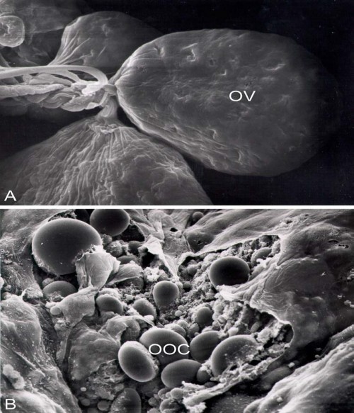 Fig.:
Fig.: Scanning electron micrograph of the ovarioles (OV) of female
Ochlerotatus rusticus. X 1 550. B.Oocytes (OOC) of female
Ochlerotatus rusticus. X 1 050.
How to cite this article:
Milagros M Greif. Studies on ovaries of mosquitoes using light and scanning microscopy. Int J Mosq Res 2016;3(3):47-50.