Studies on ovaries of mosquitoes using light and scanning microscopy
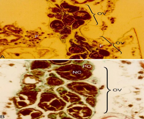
 Fig. 1:
 A Light micrograph of the ovary of
Aedes aegypti showing two clusters of ovarioles (OV) with spongy fat body (F) which surrounds the entire clusters of ovarioles of
Aedes aegypti. X2 500 B. An ovariole (OV) showing the presumptive follicle (PO) which is attached to the nurse cell (NC). X 5 000. Both after Goldner stain
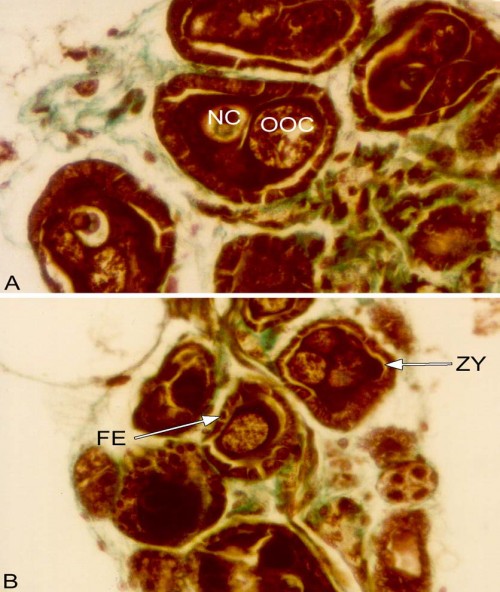
 Fig. 2:
 A. Light micrograph of an oocyte (OOC) and nurse cell (NC) of
Aedes aegypti. X 10 000. B. Zone of yolk protein uptake (ZY) in the epithelial follicle (FE) in the oocyte of
Aedes aegypti. X 7 5000. Both after Goldner stain
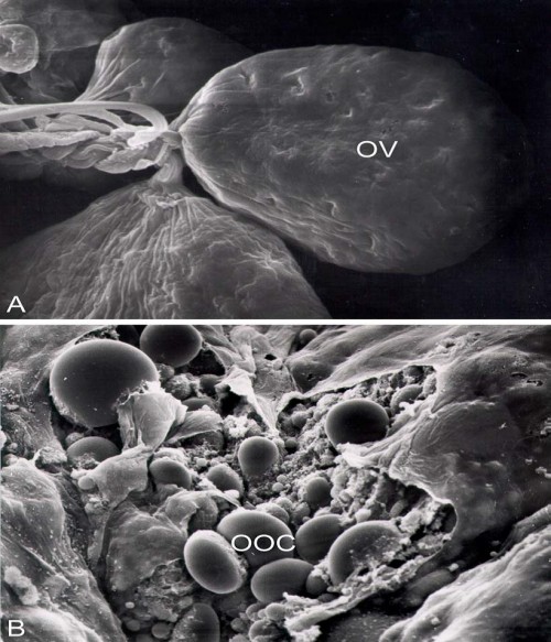
 Fig. 3:
 Scanning electron micrograph of the ovarioles (OV) of female
Ochlerotatus rusticus. X 1 550. B.Oocytes (OOC) of female
Ochlerotatus rusticus. X 1 050.


