Larvicidal activity of temephos released from new chitosan/alginate/gelatin capsules against Culex pipiens
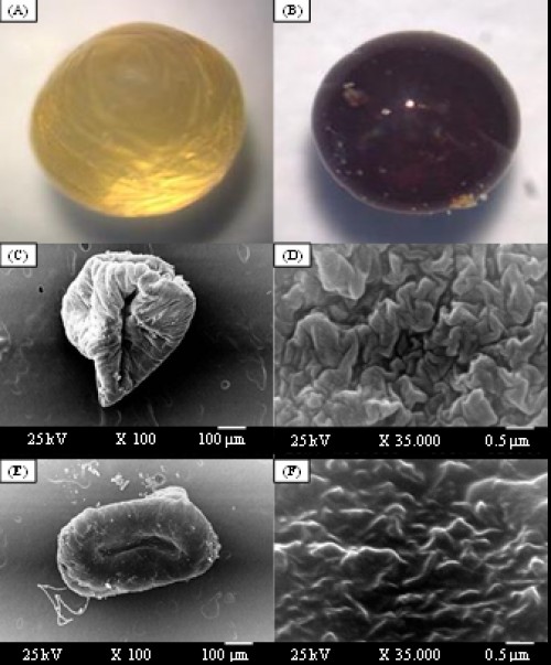 Fig. 1:
 Stereo optical microscope morphology of whole shape (magnification, X10) of the wet capsules prepared from chitosan (1%), alginate (1%), glutaraldehyde (2%) and gelatine of 2.5%, before (A) and after (B) drying. SEM photograph of unloaded capsules (C) and its surface morphology (D); and SEM photograph of loaded capsules with temephos (E) and its surface morphology (F). Scale bar 100 µm and magnification x100 for whole capsules and scale bar 0.5 µm and magnification x35000 for their surface morphologies
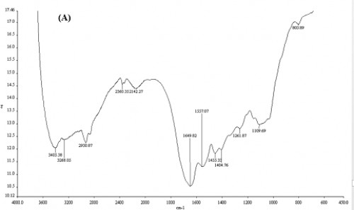 Fig. 2:
 FT-IR spectra of unloaded
(A) capsules with temephos.
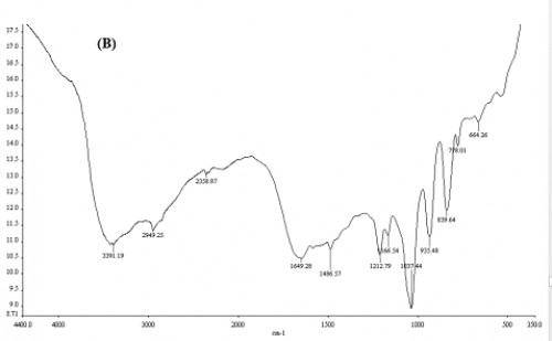 Fig. 3:
 FT-IR spectra of loaded
(B) capsules with temephos.
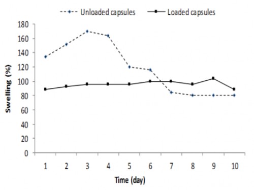 Fig. 4:
 Swelling kinetics of loaded and unloaded capsules with temephos during ten days.
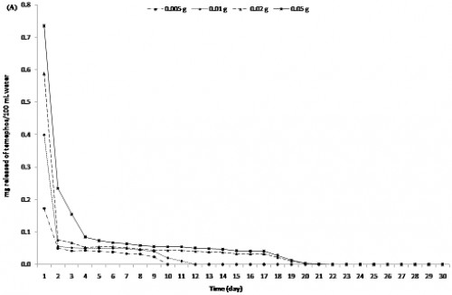 Fig. 5: In vitro
 release kinetics of the larvicide temephos by UV spectrophotometric assay in running (A) and in stagnant water (B).
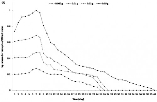 Fig. 6: In vitro
 release kinetics of the larvicide temephos by UV spectrophotometric assay in running (A) and in stagnant water (B).
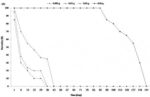 Fig. 7: In vivo
 release kinetics according to mortality percentage for different weights of capsules (0.005, 0.01, 0.02, and 0.05 g) loaded with temephos (7.35%) calculated by U.V spectroscopy for a period of time.
(A): In running water (exchange of larvae and water together) and
(B): in stagnant water (exchange of larvae only)
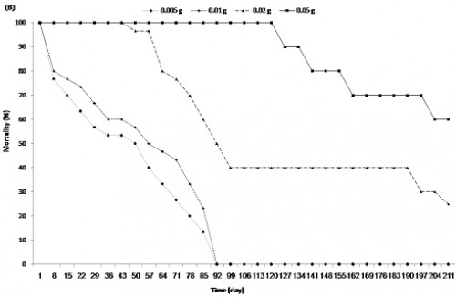 Fig. 8: In vivo
 release kinetics according to mortality percentage for different weights of capsules (0.005, 0.01, 0.02, and 0.05 g) loaded with temephos (7.35%) calculated by U.V spectroscopy for a period of time.
(A): In running water (exchange of larvae and water together) and
(B : in stagnant water (exchange of larvae only)


