- Printed Journal
- Indexed Journal
- Refereed Journal
- Peer Reviewed Journal
Subscribe Print Journal

Zoological Record Indexing

Important Information
- Helpline for Authors
- India: +91-9711224068
- Toll Free: 1800-1234070
- Working hours 10:00 AM-06:00 PM
Issue Bar
Open Access
Side Bar
Identifier

Vol. 6, Issue 4, Part A (2019)
Larvicidal potential and ultra-structural changes induced after treatment of Culex pipiens L. (Diptera: Culicidae) larvae with some botanical extracted oils
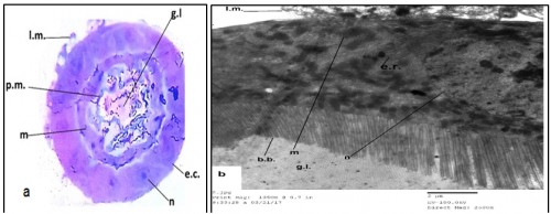
Fig. 1: Transverse section (a) (400 X) and electron micrograph (b) (4000X) in the midgut region of untreated 3rd larval instar of Culex pipiens.( e.c.= epithelial cell. g.l.= gut lumen. n= nucleus. l.m.= longitudinal muscle. b.b.= brush border, p.m. Peritrophic membrane, m =microvilli, m=mitochondria, e.r.= endoplasmic reticulum)
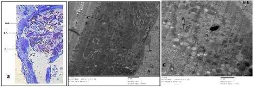
Fig. 2: Transverse section (a)(400X) and electron micrograph (b&c) (2000X & 2500X) in the midgut region of Culex pipiens larvae treated with Azadirachta indica.(e.c.= epithelial cell. g.l.= gut lumen. n= nucleus. L.m.= longitudinal muscle. b.b.= brush border. v= vacuoles).
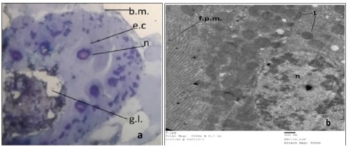
Fig. 3: Transverse section (a) (400X) and electron micrograph (b) (4000X) in the midgut of 3rd larval instar of Culex pipiens treated with Eucalyptus regnans.( e.c.= epithelial cell. g.l.= gut lumen. n= nucleus. b. m. = basement membrane. L= lipids, f.p.m = folded plasma membrane
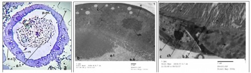
Fig. 4: Transverse section (a ) (400X) and electron micrograph (b & c ) (1000X &3000X) in the midgut of treated 3rd larval instar of Culex pipiens treated with Piper nigrum.( e.c.= epithelial cell. g.l.= gut lumen. n= nucleus. b.b.= brush border, ch= chromatin. p.m.=peritrophic membrane.
Zoological Record Indexing
Important Information
- Helpline for Authors
- India: +91-9711224068
- Toll Free: 1800-1234070
- Working hours 10:00 AM-06:00 PM
Issue Bar
Open Access
Side Bar
Identifier
Related Links

International Journal of Mosquito Research